|
Related
Documents:
M13mp18
GenBank/EMBL accession
number M77815.
Restriction
sites
Sequence
Not available from Fermentas
M13mp19
GenBank/EMBL accession
number L08821.
Sequence
Not available from
Fermentas |
Additional Information:
The M13mp18 and M13mp19 vectors are derivatives of the single-stranded,
male-specific filamentous DNA bacteriophage M13. Both vectors are 7250 bp
in length and have the same MCS inserted in opposing orientations. The vector
DNAs contain a region of E.coli operon lac containing CAP
protein binding site, promoter Plac, lac repressor binding
site and 5'-terminal part of lacZ gene encoding the N-terminal fragment
of beta-galactosidase (codons 6-7 of lacZ are replaced by MCS). This
fragment, whose synthesis can be induced by IPTG, is capable of intra-allelic
(a) complementation with a defective form of beta-galactosidase encoded by
host (mutation lacZDM15). This results in appearance of blue plaques on
media containing IPTG and X-gal. Recombinant phages containing inserts that
destroy the reading frame of lacZ are revealed as colorless plaques.
Synthesis of viral (plus) single-stranded DNA requires the phage-encoded gene
II, X and V proteins. It is initiated at ori (+) and proceeds in the direction
indicated. The conversion of plus DNA strands to double strands does not
require any of the phage genes. DNA synthesis is initiated by a 30-nucleotide
RNA primer synthesised by host's RNA polymerase and starting at ori (-).
Double-stranded circular DNA (replicative form, or RF) can be isolated from
cells by standard plasmid preparation techniques and used for cloning
experiments, while the single-stranded viral DNA (+ strand) can be isolated
from phage particles collected from culture medium.
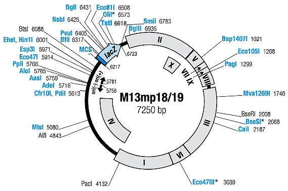
The M13mp18/19 genes are shown on the map (M13 genes are transcribed
clockwise). The map shows enzymes that cut M13mp18/19 DNA once. Enzymes
produced by Fermentas are shown in green. The coordinates refer to the
position of first nucleotide in each recognition sequence.
References
-
Norrander, J., Kempe, T. and
Messing, J., Construction of improved M13 vectors using
oligodeoxynucleotide-directed mutagenesis, Gene, 26, 101-106, 1983.
- Yanisch-Perron, C., Vieira, J. and Messing, J., Improved
M13 phage cloning vectors and host strains: nucleotide sequences of the
M13mp18 and pUC19 vectors, Gene, 33, 103-119, 1985.
Enzymes which cut M13mp18 DNA once:
AasI 5759, Acc65I 6243,
AdeI 5716,
AloI 5765,
Alw44I* 4743, BamHI 6252, BfiI 6317, BglI 6431, BglII 6935,
BseRI 2008, Bsp1407I 1021,
CaiI 2187,
Cfr9I 6247,
Cfr10I
5613, Ecl136II 6237,
Eco47I
5914, Eco81I 6508,
Eco105I
1268, EcoRI
6231, EheI
6001, Esp3I
5971, Hin1I
6001, HincII 6264,
HindIII
6282, KpnI
6243, MlsI
5080, Mva1269I 1746,
NsbI 6425,
OliI* 6573, PacI 4132, PaeI 6276, PagI 1299, PdiI 5613, PpiI 5766, PstI 6270, PvuI 6405, SacI 6237, SalI 6264, SdaI 6269, SmaI 6247, SmiI 6783, TstI 6618, XbaI 6258, XmiI 6264.
* According to our experimental
data:

Coordinates of M13mp18/19 genes (termination codons included):
I 3196-4242
II
6849-831
III 1579-2853
IV 4220-5500
V
843-1106
VI 2856-3194
VII 1108-1209
VIII
1301-1522
IX 1206-1304
X 496-831
Multiple Cloning Sites
M13mp18

M13mp19


